
A research team led by Professor Wang Kunfeng from the College of Information Science and Technology has carried out interdisciplinary medical‑engineering research in partnership with the clinical team headed by Chief Physician Ma Guolin at China‑Japan Friendship Hospital. Centering on intelligent medical image analysis and computer‑aided disease diagnosis, the joint work targets key challenges in medical image segmentation, including accurate identification of small lesions, characterization of complex tumour structures, and high‑quality segmentation under limited annotation conditions. The team keeps exploring in‑depth integration between artificial intelligence and clinical medical imaging and has secured a string of research achievements. Relevant collaborative papers have been published in international journals such as Expert Systems with Applications, IEEE Sensors Journal and Information Fusion, and two invention patents have been filed.
Medical image segmentation represents an interdisciplinary field bridging artificial intelligence and medical imaging, with critical application value in computer‑aided disease diagnosis, lesion detection, treatment planning and therapeutic effect evaluation. Nevertheless, medical images are characterised by small target scales, blurred boundaries, sophisticated structures and diverse imaging modalities. Meanwhile, high‑quality pixel‑level annotations heavily rely on professional radiologists, leaving conventional deep‑learning approaches plagued by high annotation costs, difficulty in accurate characterisation of complex lesions and limited model generalisation performance.
To tackle the above‑mentioned challenges, the research team and China‑Japan Friendship Hospital have selected clinically typical diseases including pituitary microadenoma and brain tumours as research subjects. Systematic investigations have been conducted from multiple perspectives such as spatio‑temporal feature modelling, knowledge distillation and multi‑teacher collaborative learning. A coherent research roadmap has been established, ranging from precise lesion detection and segmentation to unsupervised medical image segmentation and reliable knowledge transfer under weak‑supervision settings. It offers innovative research insights for cutting manual annotation costs for medical images and boosting the accuracy and generalisability of intelligent segmentation models.
(1) Spatio‑temporal‑feature‑integrated medical image detection and segmentation for precise diagnosis of pituitary microadenoma
Pituitary microadenoma refers to small‑sized lesions with relatively blurred boundaries. Their early detection and precise localisation are vital for clinical diagnosis and treatment. Dynamic Contrast‑Enhanced Magnetic Resonance Imaging (DCE‑MRI) can deliver dynamic enhancement information of lesion tissues changing over time. However, given the tiny size of pituitary microadenoma and limited MRI image resolution, it remains challenging to make full use of spatial and temporal information embedded in images for accurate detection and segmentation.
To address this clinical problem, the joint team has performed computer‑aided diagnosis research for pituitary microadenoma based on spatio‑temporal features. The research paper Computer‑aided diagnosis of pituitary microadenoma on dynamic contrast‑enhanced MRI based on spatio‑temporal features was published in the international journal Expert Systems with Applications in 2025.
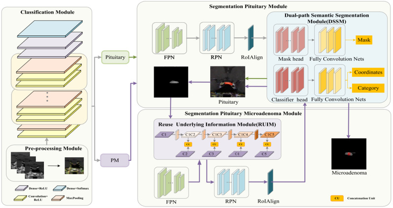
A spatio‑temporal‑information‑integrated medical image detection‑and‑segmentation model is put forward in this study. To mitigate the risk of losing fine spatial details caused by the tiny scale of pituitary microadenoma, a multi‑scale feature fusion module is designed. It extracts sufficient target semantic information while preserving distinct spatial details, enabling positive‑case identification leveraging dynamic DCE‑MRI information. For lesion segmentation, after feature alignment via ROI Align, a dual‑path semantic segmentation module is embedded into the detection network to further mine fine‑grained spatio‑temporal semantic features and enhance segmentation accuracy for the pituitary region and lesions. Moreover, a Reuse Underlying Information Module (RUIM) is devised to reuse low‑level feature information from the Feature Pyramid Network and strengthen the model’s capacity to spot small‑scale pituitary microadenoma lesions.
Experimental results demonstrate that the proposed method attains an accuracy of 97.10% and an mAP of 50.24% for computer‑aided pituitary microadenoma diagnosis, outperforming multiple representative deep‑learning models for medical images. This research provides a new technical route for intelligent detection and segmentation of pituitary microadenoma using spatio‑temporal information from DCE‑MRI and validates the clinical application potential of artificial intelligence in radiological computer‑aided diagnosis.
(2) Multi‑level knowledge distillation for medical image segmentation in scenarios without pixel‑level annotations
With the advancement of deep learning, high‑quality pixel‑level annotations serve as a cornerstone for training medical‑image‑segmentation models. For lesions such as brain tumours featuring intricate structures and variable morphology, however, producing high‑quality pixel‑level annotations demands substantial time and effort from specialist clinicians, which greatly restricts the utilisation of large‑scale medical‑image datasets.
To resolve this bottleneck, the team extended its research scope to unsupervised medical image segmentation and proposed MedDistill, a multi‑level knowledge distillation framework, in collaboration with China‑Japan Friendship Hospital. The paper MedDistill: Multi‑Level Distilled Co‑Learning for Unsupervised Medical Image Segmentation was published in IEEE Sensors Journal in 2026.
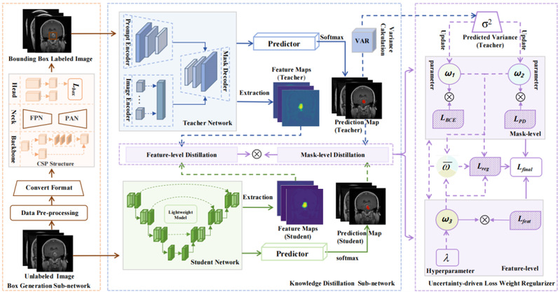
This study harnesses the powerful visual representation and segmentation capabilities of foundation vision models to generate effective supervision signals for lightweight medical‑image‑segmentation networks without pixel‑level manual annotations. First, a pre‑trained object‑detection model generates candidate tumour bounding boxes as prompts fed into the foundation vision model, which in turn yields high‑quality soft pseudo‑labels. A lightweight segmentation network acts as the student model and learns segmentation competence from the foundation vision model via knowledge distillation, accomplishing brain‑tumour segmentation free of pixel‑level manual annotations.
Different from approaches that transfer knowledge merely through final segmentation outputs, MedDistill introduces knowledge distillation at intermediate feature levels. Meanwhile, an uncertainty‑aware loss‑weight regulariser is designed to dynamically adjust weights for segmentation loss, probability distillation loss and feature distillation loss according to the reliability of diverse supervision signals. It improves knowledge‑transfer efficiency and stabilises and enhances model training robustness. Experiments on public and private brain‑tumour datasets show that MedDistill achieves competitive segmentation performance under unsupervised medical‑image‑segmentation settings and surpasses several state‑of‑the‑art existing approaches. This work corroborates the application value of collaborative learning between foundation vision models and lightweight segmentation networks in alleviating annotation dependence for medical images. It delivers new research perspectives for the paradigm shift of intelligent medical‑image analysis from reliance on extensive manual annotations towards adequate utilisation of pre‑trained models and weak‑supervision information.
(3) Uncertainty‑aware multi‑teacher knowledge distillation for weakly‑supervised medical image segmentation
Building on unsupervised‑learning research, the team further investigated unstable pseudo‑label quality under weak‑annotation conditions for medical image segmentation. In real‑world clinical workflows, clinicians can readily provide weak‑supervision information such as lesion bounding boxes or point annotations, yet obtaining fine‑grained pixel‑level segmentation annotations remains costly. Hence, it is essential to make good use of pseudo‑labels produced by foundation vision models and mitigate the adverse impact of pseudo‑label noise on model training for efficient intelligent medical‑image segmentation.
To overcome this challenge, the joint team proposed the UCM‑Distill multi‑teacher knowledge distillation framework. The paper UCM‑Distill: Multi‑Teacher Distillation with Uncertainty Modeling and Contrastive Learning for Weakly Supervised Medical Image Segmentation was published in Information Fusion in 2026.
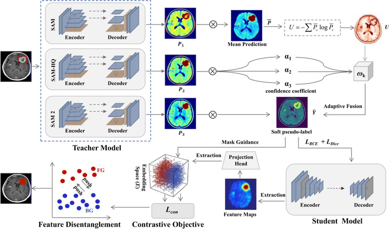
This study expands the single vision model into a multi‑teacher collaborative knowledge distillation framework, which takes full advantage of complementary strengths of different foundation vision models in medical‑image target recognition and segmentation. A consistency‑regulated uncertainty‑aware pseudo‑label generation module is devised. It fuses prediction outputs from multiple teacher models, models inter‑teacher prediction consistency and uncertainty within aggregated predictions, and modulates the contribution of each teacher’s prediction to final pseudo‑labels via continuous weighting to boost pseudo‑label reliability under weak‑supervision conditions. Additionally, a mask‑guided contrastive distillation mechanism is proposed. Pseudo‑labels are adopted to separate foreground and background regions so as to enlarge feature‑space inter‑class separability. In this way, the student network can not only learn segmentation outputs from teacher models but also acquire more discriminative feature representations.
Systematic experiments are conducted on 2D/3D public brain‑tumour datasets and a private pituitary microadenoma dataset. Experimental results verify stable segmentation performance of the proposed method across varying data scales, diverse imaging modalities and multiple segmentation tasks, accompanied by favourable cross‑dataset generalisation capability. This research further expands the application scope of knowledge distillation and foundation vision models in weakly‑supervised medical image segmentation, and offers an innovative solution for constructing high‑performance intelligent medical‑image‑analysis models with limited manual annotations.
The above three studies target pituitary microadenoma and brain‑tumour medical‑image‑segmentation tasks and follow a clear progressive research logic. The first work focuses on specific clinical diseases and imaging features, tapping spatio‑temporal contextual information embedded in DCE‑MRI to elevate detection and segmentation performance for small lesions. The second study addresses the absence of pixel‑level annotations and implements unsupervised medical‑image segmentation leveraging foundation vision models and knowledge distillation. The third study adopts multi‑teacher collaboration, uncertainty modelling and contrastive learning to explore more dependable knowledge transfer and pseudo‑label generation under weak‑supervision conditions.
From spatio‑temporal feature modelling to multi‑level knowledge distillation and further to uncertainty‑aware multi‑teacher collaborative learning, these three studies continuously broaden the application boundaries of artificial intelligence in medical image segmentation, forming a continuous research pipeline covering precise lesion detection, low‑annotation segmentation and collaborative learning with foundation vision models.
Guo Te, PhD candidate from the College of Information Science and Technology, is the first author of the three papers. Corresponding authors are Professor Wang Kunfeng from the College of Information Science and Technology and Chief Physician Ma Guolin from China‑Japan Friendship Hospital. Beijing University of Chemical Technology is designated as the first‑affiliated institution.
Moving forward, the research team will deepen scientific collaboration with China‑Japan Friendship Hospital. Future work will explore key topics in intelligent medical‑image analysis, such as low‑cost annotation, cross‑modal learning, foundation vision models, multimodal large models and generalisation in clinical scenarios. Further integration between artificial‑intelligence technologies and clinical demands will be advanced to drive the translation of medical‑image‑AI technologies from laboratory research towards dependable and practical clinical auxiliary diagnosis and treatment, and provide technical support for research and deployment of intelligent medical‑imaging technologies.